


テクニカルインフォメーション
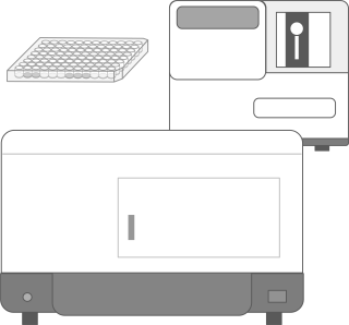
高解像度のモノクロCMOSカメラを搭載し、電動フォーカス、電動フィルター切替により、蛍光および可視光のゲルイメージングに適しています。厚みのあるアガロースゲルやワイドサイズのアクリルアミドゲルなど、様々な大きさと厚さのゲルや膜の撮影に対応しています。ゲルの切り出しに最適なガードも付属しています。
A high-resolution monochrome CMOS camera equipped with motorized focus and motorized filter switching is suitable for fluorescence and visible light gel imaging. It supports imaging of various sizes and thicknesses of gels and membranes, including thick agarose gels and wide-sized acrylamide gels. It also comes with a guard, ideal for gel cutting.
電源・撮影装置
- WSE-3100_パワーステーションギブリⅠ_使用方法_JPN
電気泳動/ブロッティング用電源装置 PowerStation GhibliⅠ パワーステーションギブリⅠ(WSE-3100)の使用方法についてご紹介します。 業界初!のタッチパネル式電源装置です。 大きなカラー液晶画面で見やすい表示と簡単操作! これ1台で電気泳動もブロッティングもOKです。
- u PAGEL H_ゲルの強度について_JPN
高い物理的強度と分離能を備えた究極のゲル『u-PAGEL H』が新登場! 新規架橋剤を用いることで、従来の低濃度ゲルよりも強度が優れ、やぶれにくいことが特長です。 染脱色操作やブロッティング操作において、ゲルの取り扱いが格段にしやくなりました。 ※強度を説明するためにゲルを引っ張っています。操作時には丁寧に扱ってください。
- PQPコントロールソフトウェア
PQPコントロールソフトウェアはペリスタクォンタムポンプ(WSP₋3300/WSP-3310)を4台までPCからコントロールできる外部制御ソフトウェアです。キャリブレーション/マニュアル送液/プログラム送液のモードがあります。プログラム送液は複数のステップを構成でき、流量勾配送液や同じ流量制御を繰り返すリピート送液などが可能です。直感操作で複雑な制御プログラムをかんたんに作成できます。
- ミニサイズゲルの作製方法_JPN
ゲル作製キット(AE-6401)を使用したミニサイズゲル(90×80mm)の作製方法をご紹介します。
- WSP-3300_ペリスタクォンタムポンプ_操作方法_JPN
現存ペリスタルティック方式ポンプ製品が未到、未実現の新技術を採用! ATTO は半世紀を超えるペリスタポンプの開発・生産・販売の経験や知見に基づき、ご要望の特に多い流量高精度をはじめとした仕様全項目を見直す改良を行い、新製品ペリスタクォンタムポンプを開発しました。
- WSE-5400 Printgraph Classic_JPN
「WSE-5400 Printgraph Classic」はカラーCMOSカメラを搭載し、高速起動~撮影~プリントのスタイルを持ちながら、画質にもこだわったゲル撮影装置です。
- WSP-3310_PeristaQuantumPumpの使用方法_JPN
- WSE-6300_LuminoGraphⅢ_化学発光撮影の方法_JPN
WSE-6300 LuminoGraphⅢは解像力の高い6M CCDカメラと世界最高峰の明るさ「F0.8」を誇るレンズを搭載したタッチパネル式ケミルミ撮影システムです。この装置を使用した化学発光撮影の方法をご紹介します。
- WSE-7010_Ezlabel FluoroNeo_使用方法
電気泳動(SDS-PAGE)用の試料調製をしながらタンパク質・ポリペプチドに蛍光標識(ラベル化)をするキットです。EzLabel FluoroNeoはタンパク質溶液と混ぜて「3分間」加熱するだけでアミノ基を蛍光標識します。タンパク質の蛍光標識をしても移動度はほとんど変わりません。また、ウエスタンブロット解析(膜上での検出、抗体との反応)にも利用可能です。
- WSE-6300_LuminoGraphⅢ_蛍光撮影の方法_JPN
WSE-6300 LuminoGraphⅢは解像力の高い6M CCDカメラと世界最高峰の明るさ「F0.8」を誇るレンズを搭載したタッチパネル式ケミルミ撮影システムです。この装置を使用した蛍光撮影の方法をご紹介します。
- ペリスタポンプ_操作方法_JPN
アトーで最も実績のあるスタンダードタイプのペリスタポンプです。クロマトグラフィー用のポンプとしてはもちろんとこと、培養液の送液や環流実験など、ラボなどの小規模から生産工場まで幅広い用途にご使用いただけます。
- WSE-1190_多連ミニスラブゲル作製器_使用方法_JPN
ミニサイズゲル(90×80mm)のポリアクリルアミドゲル作製器の紹介動画です。一度に4枚まで作製することができます。ワイドサイズ(140×80mm)、コンパクトサイズ(60x60mm)に対応した作製器もご用意しています。
- WSE-1150_パジェラン Ace_使用方法_JPN
ミニサイズゲル(90×80mm)のポリアクリルアミドゲル電気泳動槽の紹介動画です。一度に2枚まで泳動することができ、専用電源付きです。ゲルプレートを両面から恒温することで、スマイリングを低減します。
- WSL-2300_Phelios AL _微量測定方法_JPN
WSL-2300 Phelios ALは、吸光度測定(ABS)、発光測定(LUMI)、微量測定(NANO)が可能なプレートリーダーです。モノクロメーターを採用し200nm~999nmの広範囲の測定が可能です。発光測定は400nm~700nmの計測が可能です。 計測容器は、マイクロプレート(6/12/24/48/96/384)、微量測定にはオプションの「Nano Volume Plate」を用いて、2μL~の微量サンプル(DNA、RNA、タンパク質)の測定が可能です。
- WSE-6300_LuminoGraphⅢ_可視光撮影の方法_JPN
WSE-6300 LuminoGraphⅢは解像力の高い6M CCDカメラと世界最高峰の明るさ「F0.8」を誇るレンズを搭載したタッチパネル式ケミルミ撮影システムです。この装置を使用した可視光撮影の方法をご紹介します。
- WSL-2300_Phelios AL_操作方法_JPN
6/12/24/48/96/384ウェルプレート対応の吸光・発光プレートリーダーです。カイネティクスもエリアスキャンも、スペクトルもおまかせください。オプションのNano Volume plateを使用すると、たった2µLの微量試料が、一度に24検体測定できます。
- WSE-1500_ディスクラン Ace_使用方法_JPN
2次元電気泳動の1次元目(等電点)のディスクゲル電気泳動用の電源一体型電気泳動装置です。
- WSE 1030_コンパクトPAGE Neo_使用方法_JPN
コンパクトサイズ(60×60mm)のポリアクリルアミドゲル用電源一体型電気泳動装置の操作説明動画です。6種類の出力モードを搭載しているので、ゲル濃度や泳動バッファーを変えることで、SDS-PAGEやNative PAGEなど様々な電気泳動ができます。
- WSE-1040_コンパクトPAGE Neo_操作方法_JPN
コンパクトサイズ(60×60mm)のポリアクリルアミドゲル用電源一体型電気泳動装置の操作説明動画です。6種類の出力モードを搭載しているので、ゲル濃度や泳動バッファーを変えることで、SDS-PAGEやNative PAGEなど様々な電気泳動ができます。WSE-1040はツインタイプなので、ゲル1~2枚の泳動が可能です。
- WSE 1010_コンパクトPAGE Ace_使用方法_JPN
コンパクトサイズ(60×60mm)のポリアクリルアミドゲル用電源一体型電気泳動装置の紹介動画です。最速10分、標準30分で泳動できます。
- WSE 1025_コンパクトPAGE Ace twin _使用方法_JPN
コンパクトサイズゲル(60×60mm)のポリアクリルアミドゲル電気泳動槽の紹介動画です。一度に2枚まで泳動することができます。専用電源付きです。
- WSE 1165_ラピダス ミニスラブ電気泳動槽_使用方法_JPN
ミニサイズゲル(90×80mm)のポリアクリルアミドゲル電気泳動槽の紹介動画です。一度に2枚まで泳動することができます。ゲルプレートを両面から恒温することで、スマイリングを低減します。
- WSE-1710_サブマージ・ミニ_使用方法_JPN
電源搭載型のアガロースゲル電気泳動装置 サブマージ・ミニ(WSE-1710)の使用方法についてご紹介しております。
- AE-6100_サブマージ・アガロース電気泳動装置(小型)_使用方法_JPN
小型のサブマリン型アガロースゲル電気泳動装置(AE-6100)の使用方法についてご紹介しております。
- AE-6111_サブマージ・アガロース電気泳動装置(中型)_使用方法_JPN
中型のサブマリン型アガロースゲル電気泳動装置(AE-6111)の使用方法についてご紹介しております。
- AE-6500_ラピダスミニスラブ電気泳動槽_使用方法_JPN
ラピダスミニスラブ(AE-6500)の操作方法をご紹介します。
※2020年3月をもって販売終了いたしました。 - ピタットクリア_使用方法_JPN
ピタットクリアは、ゲルやメンブレンをはさみ、イメージャーで撮影する時に使用する撮影用フィルムです。
張りのあるフィルムはシワが寄らず、気泡も入りにくいのでクリアなイメージが撮影できます。はさむだけで、破れやすいアクリルアミドゲルのハンドリングが格段にUPします。 - u-PAGEL H イメージ動画
u-PAGEL H は、新規架橋剤の採用により従来のプレキャストゲルよりもポアサイズが大きく、かつ、強度の高いゲルです。従来のプレキャストゲルと比較して、泳動試料が目詰まりせずにゲル内に入るため、今まできれいに分離できなかった高分子バンド (200kDa≦) も高解像度に分離できます。u-PAGEL H は、新規架橋剤によりもたらされた効果として、優れた機械的強度を備えていることから、低濃度ゲルであっても染脱色操作、あるいは、ブロッティング操作において、ゲルがやぶれてしまうことが少なくなり、操作性が飛躍的に向上しました。
- Native sample preparation_JPN
ATTOの試薬キットを使用した、Native-PAGE用のタンパク質抽出および電気泳動サンプル調製方法をご紹介します。ATTOの試薬キットを使用した、Native-PAGE用のタンパク質抽出および電気泳動サンプル調製方法をご紹介します。
- Native-PAGE_JPN
ATTOの試薬を使用したTris系およびTris/Glysine系ゲルを使用したBlue Native PAGEとHigh Resolution Clear Native PAGEの方法をご紹介します。ATTOの試薬を使用したTris系およびTris/Glysine系ゲルを使用したBlue Native PAGEとHigh Resolution Clear Native PAGEの方法をご紹介します。
- WSC 2900_ローテーターatto_使用方法_JPN
1.5mL、15mL、50mLチューブをミキシングできるロータリーミキサー『WSC-2900 ローテーターatto』のご紹介です。
- AE-1460_EzBlot_使用方法_JPN
EzBlot (イージーブロット)は、タンパク質のセミドライ式ブロッティング用の3溶液系ブロッティング溶液です。 高効率な転写が可能です。
- WSE-4057_QBlot kit M_使用方法_JPN
トランスファーパック QBlot kit M(WSE-4057)の使用方法を動画でご紹介します。
- WSC-2800_マイミニボルテックス_使用方法_JPN
小型で省スペースながら、50mLチューブも攪拌可能! パワフルなボルテックスミキサー『WSC-2800 MyMiniVortex』のご紹介です。
- ウエスタンブロッティングの操作方法_JPN
アトーの装置や試薬を使用したウエスタンブロッティングの操作方法をご紹介します。
- WSC-2615_マイミニブロック C&H_使用方法_JPN
手のひらサイズのブロックインキュベータです。 0-100℃の温度設定、0.2mLチューブから50mLチューブまで自由自在。 小さくて頼りになる相棒です。
- WSC-2700_マイミニスピン_使用方法_JPN
小型遠心機 MyMiniSpin マイミニスピン(WSC-2700)の使用方法についてご紹介します。 4種類のテストチューブを同時に遠心可能! 便利なタイマー機能付き!
- WSE-7160_EzStainAQua MEM_How to use_ENG
※英語字幕の動画です。
ブロッティング膜上のタンパク質を検出するCBB染色・脱色キットです。脱色後は抗体反応が可能で、トータルタンパク質によるノーマライズにも使用できます。 - u PAGEL H_セミドライブロッティングのコツ_JPN
高い物理的強度と分離能を備えた究極のゲル『u-PAGEL H』が新登場! 低濃度ゲルは、高濃度ゲルに比べるとやわらかいため、取り扱いが難しいです。 そこでセミドライブロッティングをする際のコツについてご紹介します。
- WSF-2000MH_ファーモグラフⅢ_使用方法_JPN
ファーモグラフは、微生物発酵などによって発生するガス量の計測装置です。ファーモグラフIIIは「日本イースト工業会のパン用酵母試験法」で採択されたファーモグラフの後継機種です。
最大20点までの試料のガス発生量を同時並行で自動測定できる装置で、微生物酵母などの発酵能力や培地/生地組成、発酵条件を評価して育種・開発・品管等にご利用できます。 - u PAGEL H_ゲルの強度について_JPN
高い物理的強度と分離能を備えた究極のゲル『u-PAGEL H』が新登場! 新規架橋剤を用いることで、従来の低濃度ゲルよりも強度が優れ、やぶれにくいことが特長です。 染脱色操作やブロッティング操作において、ゲルの取り扱いが格段にしやくなりました。 ※強度を説明するためにゲルを引っ張っています。操作時には丁寧に扱ってください。
- WSL-1800_CytoWatcher_使用方法_JPN
WSL-1800 CytoWatcherは小型のデジタル顕微鏡です。コンパクトなボディサイズで、防湿構造なのでCO2インキュベーター内にも設置でき、培養しながらのタイムラプス撮影にも対応しています。
- WSL-1850_Wound healing captured by CytoWatcher II
コンフルエントのNIH3T3 細胞にチップでひっかき傷(創傷)を作り、修復される様子をCytoWatcher Ⅱにより10 分間隔でインターバル撮影しました。細胞が躍動的に移
動する様子が観察できます。 - WSL-1850_CytoWatcher Ⅱの使用方法_JPN
CytoWatcherⅡ/CytoWatcherⅡ FL は、生きたままの細胞や組織を、ダメージを与えることなく、長期間にわたって撮影できるデジタル顕微鏡です。省スペース、省電力設計なので、CO2インキュベータ内にコンパクトに収まり、環境温度への影響を最小限に抑えます。明視野撮影はもちろんのこと、蛍光撮影にも対応しており、再現性抜群のカラー撮影は組織切片や生体組織などの観察にも適しています。
- WSL-1850_Fluorescent Imaging of GFP expressed NIH3T3 cells
EGFP 発現ベクターを導入してから20 時間後のNIH3T3 細胞を、CytoWatcher Ⅱ FL で撮影したイメージを示しています。遺伝子導入後、18 ~ 30 時間後を20分間隔でインターバル撮影により撮影しました。
- WSL-1800_CytoWatcher_Time-lapse cell imaging of GFP cells (FL/BF merged)
※英語字幕の動画です。
Fluorescence and bright field time-lapse images of NIH 3T3 cells (a 35 mm glass bottom dish) for 48 h after transfection of EGFP expression vector, using a fluorescent imaging model of CytoWatcher. ATTO CytoWatcher. - AE-2550_Kronos Dio_Operation guide_ENG
※英語字幕の動画です。
- WSL-1800_CytoWatcher_Time-lapse cell imaging of GFP cells
Fluorescence time-lapse images of NIH 3T3 cells (a 35 mm glass bottom dish) for 48 h after transfection of EGFP expression vector, using a fluorescent imaging model of CytoWatcher. ATTO CytoWatcher.
- AB-3000_Cellgraph_Monitoring of wound healing assay with luminescence in living cell_ENG
※英語字幕の動画です
The result of the wound healing assay observed with Cellgraph are shown in this movie. NIH3T3 cells stably expressing luciferase were cultured until fully confluent. Wounds were created by scraping monolayer cells with a sterile pipette tip. Then wound closure was monitored by Cellgraph. - WSL-1800_CytoWatcher_Time-lapse cell imaging of IVF bovine embryos
Time-lapse live cell imaging system, CytoWatcher (ATTO) Time-lapse monitoring bovine embryos 6 hours after in vitro fertilization for 9 days (0.5 h-intervals) in an incubator By courtesy of Kei Imai, Department of Sustainable Agriculture, Rakuno Gakuen University, Japan ATTO CytoWatcher.
- AB-3000_Cellgraph_Quantitative Data analysis of luminescent intensities in living cells_ENG
※英語字幕の動画です。
Cellgraph Viewer is an image analysis software. It is very simple and easy to use, and has multiple function such as luminescent intensity measurement, making movie and montage... etc.. The ROI mode in analysis tools provides the function to measure bioluminescent intensities individually in any given region of interest. The result of analyzing bioluminescence intensity data can be exported as CSV format files. - WSL-1800_CytoWatcher_Time-lapse cell imaging of Wound healing assay
Time-lapse live cell imaging system, CytoWatcher (ATTO) Wound healing assay with fibroblast NIH3T3 cells for 24 h (30 minutes-intervals) in a CO2 incubator ATTO CytoWatcher.
- AB-3000_Cellgraph_Real time monitoring assay of luminescence in living cells_ENG
※英語字幕の動画です。
Bioluminescent images are acquired from NIH3T3 cells expressing SV40 promoter fused luciferase with cellgraph. The merge images of bright field and bioluminescence shown as pseudo color. - Time-lapse cell imaging, NIH3T3 cells by CytoWatcher
Time-lapse live cell imaging system, CytoWatcher (ATTO)
Growth of fibroblast NIH3T3 cells for 72 h (30 minutes-intervals) in a CO2 incubator - AB-3000_Cellgraph_Visualization of ATP oscillation with luciferase in living cell_ENG
※英語字幕の動画です。
Cellular condensation in embryonic limbs that occurs in the early stage of chondrogenesis is considered to play a critical role in the secretion of adhesion molecules and extracellular matrixes. The movie shows the visualization of ATP oscillation after the induction of chondrogenesis in ATCD5 cells transfected with an ATP-dependent Phyxothrix hirtus luciferase gene. As demonstrated in this study, the Cellgraph system is an effective.
- AB-3000_Cellgraph_Visualization of intracellular protein trafficking in living cells_ENG
※英語字幕の動画です。
Visualization of nucleocytoplasmic shuttling of importin α by the Cellgraph system. In this study, importin α gene fused with luciferase was expressed in NIH3T3 cells. The time-lapse images were acquired using three minutes exposure time at intervals of four minutes with a 40x objective lens without binning. The luminescence signal was initially detected in the cytosol, then in the nucleus. After that, the luminescence signal in the nucleus gradually increased. As shown above, the Cellgraph system is an ideal tool for observing biological events such as trafficking of proteins that occur over a prolonged period of time.
Reference: Y. Nakajima, PLoS One, 5(4), e10011 (2010) [PubMed] - AB-3000_Cellgraph_Bioluminescence imaging in brain tissue sample_ENG
※英語字幕の動画です。
Using the Cellgraph system, a brain tissue slice from a transgenic mouse that express luciferase under control of the clock gene promoter were analyzed. The Brain was removed and sectioned into 100 µm thick slices using a Microslicer, each of which was then placed in a culture insert. The time-lapse images of an SCN section acquired over a period of five days using the Cellgraph system. Using the grid measurement function of “Cellgraph Viewer”, the bioluminescence intensity in each area was analyzed and quantified. - AB-3000_Cellgraph_Bioluminescent imaging of activated GnRH neurons by kisspeptin stimulation
※英語字幕の動画です。
Ultradian rhythms of gonadotropin-releasing hormone (GnRH) gene transcription in single GnRH neurons were monitored using cultured hypothalamic slice prepared from transgenic mouse expressing a GnRH promoter-driven destabilized liciferase reporter. It was demonstrated that GnRH gene transcription was synchronously activated after kisspeptin was added to the media.
Reference: HK. Choe, Proc. Natl. Acad. Sci. U.S.A., 110(14), 5677-5682 (2013) [PubMed]
Power supply / Imaging system
- WSE-3100_PowerStation GhibliⅠ_How to use_ENG
The World’s first Touch panel control!
Simple operation with big touch color screen
Operation system with a latex glove
Broad output range
Voltage 0~500V , Current 0~3000mA
Available with High-speed mode electrophoresis and High-speed mode blotting. - u-PAGEL H_Amazing flexibility_ENG
The ultimate gel "u-PAGEL H" with amazing physical strength and separability is now available!
By using a new crosslinker, the new gel is stronger and harder to tear than conventional low-concentration gels.
The gel is easy to handle during the operations of staining, bleaching and blotting.
※The gel is pulled to explain the strength, so please handle with care when operating. - How to make an SDS-PAGE gel_ENG
This video explains how to make an SDS-PAGE gel using hand gel casting kit(AE-6401).
- WSP-3300_PeristaQuantumPump_Operation guide_ENG
Digital new peristaltic pump Improved stability, reproducibility, chemical resistance, flow rate, operability, and external control functions.
■ Metal non-contact: Bio-Safe aseptic, contamination-free, sanitary and clean liquid feed
■ Accomplish the "High Flow Accuracy" by "Quantitative flow stability" and "High flow reproducibility"
■ Low pulsation: by "12-roller rotating roller system"
■ Includes "PeristaQ dedicated standard tube"
■ Easy to use by "Touch Panel Operation and Settings"
■ Productivity improved by external control system - WSP-3310_How to use PeristaQuantumPump_ENG
- WSE-7010_Ezlabel FluoroNeo_How to use
EzLabel FluoroNeo is a kit for SDS sample preparation and simultaneously for fluorescent labeling of protein and polypeptide. Hardly to detect any differences in protein band mobility after labeling reaction. The observed gel can proceed to western blotting as usual. After blotting, transferred proteins on membrane can be detected by excitation light..
- Perista pump_Operation guide_ENG
- Standard Perista Pump
- Pumping fluids/solutions in chromatography
- Pumping fermentation broth
- Pumping chemicals in recirculation experiment
- A wide range of application, from laboratory to plant, etc
- WSE-1190_Multi Mini-Slab Gel Cast_How to use_ENG
Up to 4 pieces of Mini-Slab size (90 x 83 mm, thickness 1 mm) gels can be made simultaneously with this gel casting kit.
- WSE-1150_PageRunAce_How to use_ENG
WSE-1150 PageRunAce is a mini-slab size PAGE system with a built-in power supply.
- WSL-2300_Phelios AL _Micro-volume measurement_ENG
Only you need is 2μL 24 samples at a time In order to carry out experiments with high accuracy and reproducibility, it is necessary to measure the concentration of nucleic acids and proteins. However, the samples are limited and valuable and should be measured with minimal quantity and accuracy. The Phelios AL allows you to measure 24 samples at a time with a 2 μL sample volume using the “Nano Volume Plate” (optional). In preset mode, in addition to measuring the quantity, the spectrum for checking the purity of DNA / RNA can be automatically measured at the same time. The measured value is displayed as the average value measured three times.
- WSL-2300_Phelios AL_Measuring ABS_ENG
WSL-2300 Phelios AL is a multimode microplate reader for measuring absorbance (ABS), luminescence (LUMI), and microvolume samples (NANO: 2µL). Phelios AL enables the use of a wide range of applications and is also superior in terms of performance and economy.
- WSE-1500_DiscRun_How to use_ENG
WSE-1500 DiscRun can do 1-D electrophoresis(IEF) for 2-D PAGE system.Two-dimensional electrophoresis can be done easily with the use of a version with a power source (electricity conditions entered) and “agarGEL” precast gel and a high level of reproducibility can be obtained 150-210 min.
- WSE 1030_CompactPAGE Neo_How to use_ENG
Compact PAGE Neo is a compact size gel electrophoresis system
built-in power supply for compact size (60 x 60 mm) polyacrylamide gels. It is equipped with six types of output modes, so various electrophoresis such as SDS-PAGE and Native PAGE can be performed by changing the gel concentration and electrophoresis buffer. - WSE 1040_CompactPAGE Neo_How to use_ENG
Compact PAGE Neo is a compact size gel electrophoresis system
built-in power supply for compact size (60 x 60 mm) polyacrylamide gels. It is equipped with six types of output modes, so various electrophoresis such as SDS-PAGE and Native PAGE can be performed by changing the gel concentration and electrophoresis buffer. - WSE 1010_CompactPAGE Ace_How to use_ENG
WSE-1010 cPAGE Ace is a compact-slab size PAGE system with a built-in power supply. The gel size(60mm x 60mm) and the apparatus body are very compact and easy-to-handle.
- WSE 1025_CompactPAGE Ace twin _How to use_ENG
WSE-1025 CompactPAGE Ace Twin is a compact-slab size PAGE system with a built-in power supply. The gel size(60mm x 60mm) and the apparatus body are very compact and easy-to-handle.Up to 2 gels can be done electrophoresis.
- WSE-1165_Mini-Slab_How to use_ENG
This video explains how to use WSE-1165 Mini-Slab.
- WSE-1710_Submerge-Mini_How to use_ENG
Submarine electrophoresis system with built-in power supply! Submerge-Mini is available Hi-Speed(High voltage150V) electrophoresis, and has alarm timer function.
- AE-6100_Agarose EP kit(small type)_How to use_ENG
Agarose EP Kit (small type)is an electrophoresis system for submerged, horizontal agarose gel.
- AE-6111_Agarose EP kit(wide type)_How to use_ENG
Agarose EP Kit(Wide type) is an electrophoresis system with buffer recirculation for submerged, horizontal agarose gel.
- AE-6500_Dual Mini Slab_How to use_ENG
The AE 6500 was a best-selling model, but it has been redesigned to the AE-1165 and optimized for faster, higher resolution electrophoresis.
- WSC-2900_Rotator atto_How to use_ENG
Tabletop small volume rotary mixer "WSC-2900 Rotator atto" .
Mixing of 1.5mL, 15mL and 50mL tubes are available. - AE-1460_EzBlot_How to use_ENG
EzBlot is a transfer buffer comprised of three reagent for semi-dry blotting of protein sample.
- WSE-4057_QBlot kit M_How to use_ENG
QBlot kit series are transfer pack. Needless preparation! Convenient and easy to use.
- WSC-2800_MyMiniVortex_How to use_ENG
Compact vortex mixer "WSC-2800 MyMiniVortex".
It's compact size that fits in the palm of your hand and mixing of 1.5mL~50mL tubes are available! - Western blotting method_ENG
This video explains how to operate Western blotting using ATTO products.
- WSC-2615_MyMiniBLOCK C&H_How to use_ENG
A personal-sized compact block incubator. Temperature setting from 0 to 100 ℃, available for 0.2mL tube to 50mL tube. Convenient and easy to use.
- WSC-2700_MyMiniSpin_How to use_ENG
This video explains how to use MyMiniSpin (WSC-2700)
- WSE-7160_EzStainAQua MEM_How to use_ENG
Total protein stain kit for transfer membrane. visualization of protein in transfer membrane. check transfer unevenness before antibody reaction.
- u-PAGEL H_How to handle the gel for western blotting_ENG
The ultimate gel "u-PAGEL H" with amazing physical strength and separability is now available! The low concentration gels are softer than high concentration gels,so it's difficult for handling. How to handle the gel for western blotting!
- u-PAGEL H_Amazing flexibility_ENG
The ultimate gel "u-PAGEL H" with amazing physical strength and separability is now available!
By using a new crosslinker, the new gel is stronger and harder to tear than conventional low-concentration gels.
The gel is easy to handle during the operations of staining, bleaching and blotting.
※The gel is pulled to explain the strength, so please handle with care when operating. - WSL-1850_Wound healing captured by CytoWatcher II
- WSL-1850_CytoWatcher Ⅱ_How to use_ENG
CytoWatcherⅡ/CytoWatcherⅡ FL is a digital microscope that allows for the long-term imaging of live cells and tissues without causing damage. Designed to be space-efficient and energy-saving, it fits compactly inside a CO2 incubator, minimizing the impact on environmental temperature. It supports not only bright-field imaging but also fluorescence imaging, and its outstanding color reproduction is suitable for observing tissue sections and living tissues.
- WSL-1850_Fluorescent Imaging of GFP expressed NIH3T3 cells
- WSL-1800_CytoWatcher_Time-lapse cell imaging of GFP cells (FL/BF merged)
Fluorescence and bright field time-lapse images of NIH 3T3 cells (a 35 mm glass bottom dish) for 48 h after transfection of EGFP expression vector, using a fluorescent imaging model of CytoWatcher. ATTO CytoWatcher.
- AE-2550_Kronos Dio_Operation guide_ENG
- WSL-1800_CytoWatcher_Time-lapse cell imaging of GFP cells
Fluorescence time-lapse images of NIH 3T3 cells (a 35 mm glass bottom dish) for 48 h after transfection of EGFP expression vector, using a fluorescent imaging model of CytoWatcher. ATTO CytoWatcher.
- AB-3000_Cellgraph_Monitoring of wound healing assay with luminescence in living cell_ENG
The result of the wound healing assay observed with Cellgraph are shown in this movie. NIH3T3 cells stably expressing luciferase were cultured until fully confluent. Wounds were created by scraping monolayer cells with a sterile pipette tip. Then wound closure was monitored by Cellgraph.
- WSL-1800_CytoWatcher_Time-lapse cell imaging of IVF bovine embryos
Time-lapse live cell imaging system, CytoWatcher (ATTO) Time-lapse monitoring bovine embryos 6 hours after in vitro fertilization for 9 days (0.5 h-intervals) in an incubator By courtesy of Kei Imai, Department of Sustainable Agriculture, Rakuno Gakuen University, Japan ATTO CytoWatcher.
- AB-3000_Cellgraph_Quantitative Data analysis of luminescent intensities in living cells_ENG
Cellgraph Viewer is an image analysis software. It is very simple and easy to use, and has multiple function such as luminescent intensity measurement, making movie and montage... etc.. The ROI mode in analysis tools provides the function to measure bioluminescent intensities individually in any given region of interest. The result of analyzing bioluminescence intensity data can be exported as CSV format files.
- wsl-1800-cytowatcher-time-lapse-cell-imaging-of-wound-healing-assay
Time-lapse live cell imaging system, CytoWatcher (ATTO) Wound healing assay with fibroblast NIH3T3 cells for 24 h (30 minutes-intervals) in a CO2 incubator ATTO CytoWatcher.
- AB-3000_Cellgraph_Real time monitoring assay of luminescence in living cells_ENG
Bioluminescent images are acquired from NIH3T3 cells expressing SV40 promoter fused luciferase with cellgraph. The merge images of bright field and bioluminescence shown as pseudo color.
- Time-lapse cell imaging, NIH3T3 cells by CytoWatcher
Time-lapse live cell imaging system, CytoWatcher (ATTO)
Growth of fibroblast NIH3T3 cells for 72 h (30 minutes-intervals) in a CO2 incubator - AB-3000_Cellgraph_Visualization of ATP oscillation with luciferase in living cell_ENG
Cellular condensation in embryonic limbs that occurs in the early stage of chondrogenesis is considered to play a critical role in the secretion of adhesion molecules and extracellular matrixes. The movie shows the visualization of ATP oscillation after the induction of chondrogenesis in ATCD5 cells transfected with an ATP-dependent Phyxothrix hirtus luciferase gene. As demonstrated in this study, the Cellgraph system is an effective.
- AB-3000_Cellgraph_Visualization of intracellular protein trafficking in living cells_ENG
Visualization of nucleocytoplasmic shuttling of importin α by the Cellgraph system. In this study, importin α gene fused with luciferase was expressed in NIH3T3 cells. The time-lapse images were acquired using three minutes exposure time at intervals of four minutes with a 40x objective lens without binning. The luminescence signal was initially detected in the cytosol, then in the nucleus. After that, the luminescence signal in the nucleus gradually increased. As shown above, the Cellgraph system is an ideal tool for observing biological events such as trafficking of proteins that occur over a prolonged period of time.
Reference: Y. Nakajima, PLoS One, 5(4), e10011 (2010) [PubMed] - AB-3000_Cellgraph_Bioluminescence imaging in brain tissue sample_ENG
Using the Cellgraph system, a brain tissue slice from a transgenic mouse that express luciferase under control of the clock gene promoter were analyzed. The Brain was removed and sectioned into 100 µm thick slices using a Microslicer, each of which was then placed in a culture insert. The time-lapse images of an SCN section acquired over a period of five days using the Cellgraph system. Using the grid measurement function of “Cellgraph Viewer”, the bioluminescence intensity in each area was analyzed and quantified.
- AB-3000_Cellgraph_Bioluminescent imaging of activated GnRH neurons by kisspeptin stimulation
Ultradian rhythms of gonadotropin-releasing hormone (GnRH) gene transcription in single GnRH neurons were monitored using cultured hypothalamic slice prepared from transgenic mouse expressing a GnRH promoter-driven destabilized liciferase reporter. It was demonstrated that GnRH gene transcription was synchronously activated after kisspeptin was added to the media.
Reference: HK. Choe, Proc. Natl. Acad. Sci. U.S.A., 110(14), 5677-5682 (2013) [PubMed]